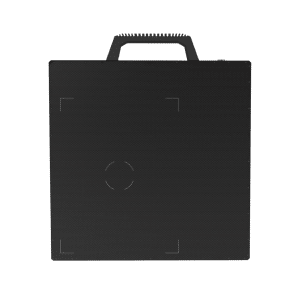
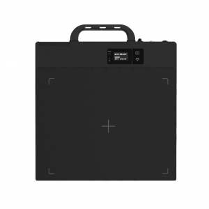
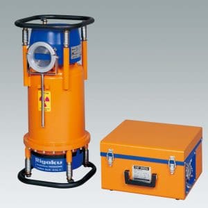
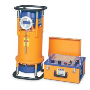
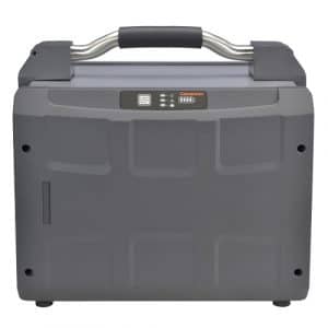
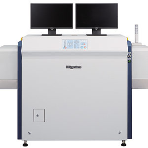
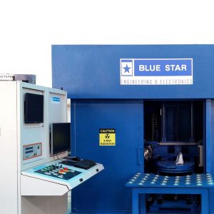
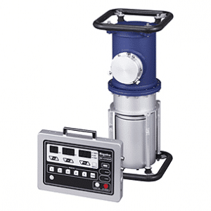
High resolution 3D CT
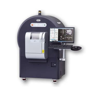
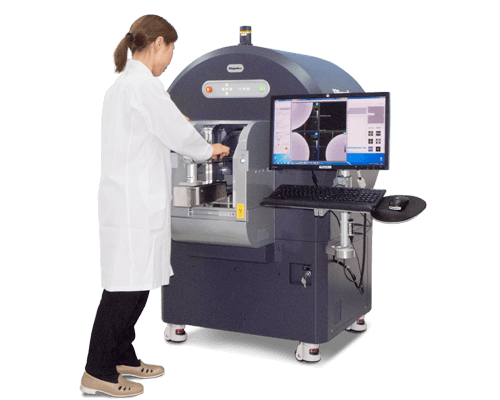
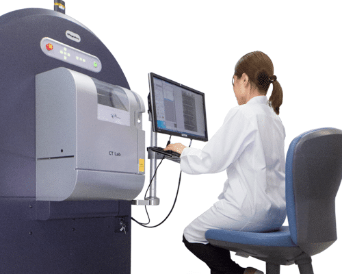
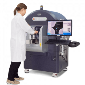
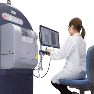
The CT Lab pursues high-speed, high-resolution, and quick sample-mounting in order to be used for R&D purposes as well as at production sites. The field-of-view and resolution can be selected at will to observe even fine structures. The maximum number of pixels is 8000×8000. High-definition 3D observation is possible at the highest level compared with other products in the same class. The maximum diameter of a measurement subject is 72 mm×36 mm. After the wide field-of-view imaging, the image of fine structure can be reconstructed in details by specifying the ROI area in the newly incorporated “wide view imaging function”.
Live mode for in-situ imaging
A 2D high-resolution Live mode with recording function is integrated to allow usage as an even faster computed radiography. The distinctive feature of this mode is that the structural change of the subject can be observed under in-situ in real time. This feature is effective for observing the change in battery cells during charge and discharge, status of cooling water hose when liquid flows through, or for measurement of structural change while heating. The observation result can be recorded so the scene when the change occurred can be checked afterwards. Note that since the power supply cable and hoses of the measurement subject can be wired outside the equipment through the insertion port on the side area, the power of the measurement subject can be turned on and off even while the measurement is in progress.
Two variations: GX90 and GX130
Two models are available: the low-powered CT Lab GX90, suited for observation of subjects such as resins, and the high-powered CT Lab GX130, suited for subjects more difficult to transmit X-ray beams such as metals.
Low running cost
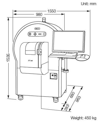
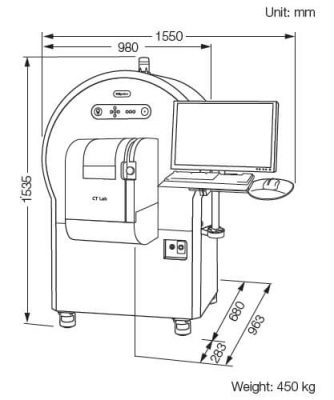
The equipment operates by 100V power supply, and cooling water is unnecessary. The equipment has casters to make it easy to move within factories and warehouses. X-ray radiation safety officer is unnecessary to operate this equipment.
CT Lab specifications
| 유형 | GX 90 | GX130 | |
| X-ray source | Tube current | ~90 kV | ~130 kV |
| Maximum power | ~200 μA ( 8W) | ~300 μA ( 39 W) | |
| Detector | 유형 | Flat panel detector | |
| CT gantry | Bore size | 613 mmΦ (Max.) | |
| Scanable range | 240 mmΦ (Max.) | ||
| CT image | Field of view | 72 mmΦ (Max.) | |
| CT image reconstruction |
15 sec. (Min.) | ||
| Resolution | 45 μm (Min.) | ||
| Number of pixels | 512 x 512 ~ 8000 x 8000 | ||
| Live image | Movie | 600 fps (Max.) | |
| Photo | 16.7 msec. (Min.) | ||
| CPU | OS | Window 7 | |
| Memory | 32 GB | ||
| HDD | 500 GB + 2 TB | ||
| Image analysis |
2D | Movie, still image, recording function | |
| 3D | Image measurement function Volume rendering function 3 surface cross section view function |
||
| Installation condition |
Power supply | 100 – 120 VAC, 1 phase, 15 A 200-240 VAC, 1phase, 8A |
|
Specifications and appearance are subject to change without notice.
CT Lab applications
Ultra-high-speed CT scan and image reconstruction
CT image can be created by CT scan in 8 seconds and reconstruction in 15 seconds at top speed. Even in the high-resolution mode, scan time of 57 minutes is achieved, which dramatically reduces the scan time which requires two to three hours for high resolution images in general.
High-resolution wide field-of-view measurement
Fine structures can be observed by selecting a field-of-view and resolution in the included original software. The minimum resolution is 4.5μm, and the maximum number of pixels is 8000×8000. In general, the field-of-view will narrow when the resolution is increased, but in CT Lab GX, the measurement can be performed while keeping the wide field-of-view even in high resolution mode.
Incorporating the “Sample-Stationary Method”
CT scan can be performed by just placing the measurement subject within the equipment, with no need to move the measurement subject. The sample can be positioned easily and CT scan can be done even for materials within a liquid with the lid kept open.
