What is X-ray microscopy?
Tomography is the study of the three dimensional structure of an object by slicing it into thin sections. Microtomography implies that the slices are very thin; thin enough to be viewed by an optical microscope. Classical tomography is a tedious and time-consuming process, and can also result in significant perturbations to the sample. In X-ray tomography, the entire sample is imaged at multiple rotation angles. This multitude of images is processed by sophisticated computer algorithms to provide a three dimensional reconstruction that can be sliced in any direction, providing new insights into the internal features of the object. X-ray microscopy provides this visualization at a resolution better than a micrometer (μm).
High-resolution, high-contrast X-ray microscope
The new nano3DX allows you see into many types of samples, including those that have low absorption contrast, for example CFRP, or denser materials like ceramic composites. The nano3DX allows you to achieve this by providing the ability to change the X-ray wavelength to enhance contrast or penetration.
Features
- Ultra-wide field of view, 25X larger volume than comparable systems
- 3 X-ray wavelengths (Cr, Cu and Mo Ka) to optimize imaging for different sample matrices
- Parallel beam geometry for high contrast and rapid data collection
- Auto 5-axis (XYZ and rotation) stage and on-axis imaging system
- High resolution three dimensional (3D) images
- High power rotating anode X-ray source
- High contrast for low-Z materials
- High-resolution CCD imager
Specs
- X-ray microscope
- Microtomography of large samples at high resolution
- X-ray generator: MicroMax-007 HF
- Tube voltage: 20 to 50 kV
- Tube current: up to 30 mA
- Target: Cr, Cu and Mo
- Detector: X-ray CCD camera
- Number of pixels: 3300 × 2500
- Pixel size: 0.27 to 4 μm
- Field of view: 0.9 × 0.7 mm to 14 × 10 mm
- Dynamic range: 16 bit
- Sample stage: automatic 5-axis stage
- Stage rotation axis accuracy: <0.5 μm
Examples
Mouse fibula at high resolution |
|
| The nano3DX is ideally suited to the analysis of bone samples at submicron resolution. One of the important features of the nano3DX is the ability to look at large samples at high resolution. In the example below, an entire mouse fibula was analyzed with the nano3DX, the first time a whole bone analysis has been performed. | |
 |
|
| Parallel beam geometry and the ability of the nano3DX to obtain rapid high-contrast images allowed for the collection of the full tomogram of the bone from the proximal to distal ends. With each slice, the structures in the bone are clearly visible, including the softer cartilage at the proximal end. The scale bars are 50 μm long in each of the insets and the bone itself is approximately 12 mm long. The bone marrow, microvessels and osteocyte lacunae are clearly depicted. | |
Pharmaceuticals |
|
| Below is shown the analysis of a single pharmaceutical particle from a capsule. The first image is a single slice through the reconstruction. The second image is an interior tomogram of the particle at 0.54 μm/voxel. The last image displays a rendering of an analysis of the pores in the sample, color coded by size (red largest, green smallest). | |
 |
|
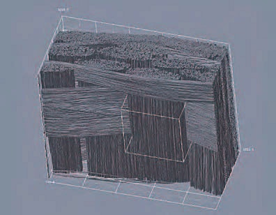
Ultra wide field of viewAt right is shown an example of a carbon fiber reinforced polymer (CFRP). The fibers are 7 μm in diameter. The image is 1.8 mm x 1.8 mm by 1.4 mm with a voxel size of 0.54 μm. The volume is represented by 3300 x 3300 x 2500 voxels. This volume is 25X larger than the measurable volume from a single scan from other systems at this resolution in a comparable time frame. |
 |
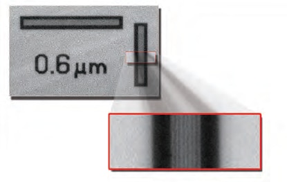
High resolutionThe two dimensional (2D) resolving power is shown directly in the images above with a transmission image of a test pattern at 0.27 μm per pixel in which lines at 0.6 μm are resolved. |
|
